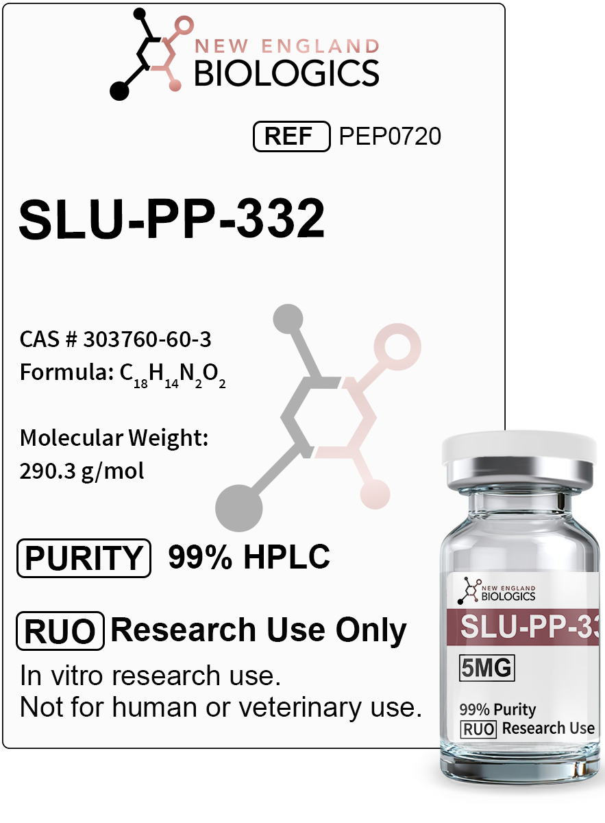
Exercise Performance and Muscle Function
Studies demonstrate that SLU-PP-332 significantly enhances physical performance through ERRα-dependent mechanisms. Research published in ACS Chemical Biology showed that sedentary mice treated with SLU-PP-332 for 7 days displayed approximately 70% longer running duration and 45% greater running distance compared to vehicle-treated controls during exhaustion testing. This exercise-mimetic effect was completely dependent on ERRα, as mice with muscle-specific deletion of ERRα showed no improvement in exercise endurance with SLU-PP-332 treatment.
The compound induces substantial changes in skeletal muscle composition and oxidative capacity. Treatment with SLU-PP-332 increases type IIa oxidative muscle fibers while enhancing succinate dehydrogenase (SDH) activity, a marker of oxidative metabolism. Histological analysis reveals increased mitochondrial content in skeletal muscle tissue, confirmed by elevated mitochondrial DNA levels relative to nuclear DNA. Expression of oxidative phosphorylation complex proteins, including complex I (NDUFB8), complex V (ATP5A), and cytochrome c, increases significantly with SLU-PP-332 administration.
Molecular studies show that SLU-PP-332 activates an acute aerobic exercise genetic program characterized by transient upregulation of DDIT4 and SLC25A25 (ATP-Mg2+/Pi mitochondrial transporter). The magnitude and temporal pattern of gene induction with SLU-PP-332 closely mirrors that observed with actual aerobic exercise, with peak expression at 1-3 hours post-treatment followed by return to baseline. RNA sequencing studies identified 442 differentially expressed genes in quadriceps muscle and 238 in gastrocnemius muscle following SLU-PP-332 treatment, with significant overlap between the compound-induced changes and genes activated by acute aerobic exercise in both mice and humans.
In separate studies examining grip strength, mice treated with SLU-PP-332 for two weeks showed measurable increases in muscle strength compared to vehicle-treated controls. The compound's effects on muscle function occur through multiple mechanisms including enhanced mitochondrial respiration in skeletal muscle cells, increased expression of oxidative metabolism genes, and promotion of muscle fiber adaptations associated with endurance capacity. Electron microscopy of muscle tissue from treated animals reveals increased mitochondrial abundance and improved mitochondrial structure compared to controls.
Sources:
Metabolic Health and Fat Loss
SLU-PP-332 demonstrates significant metabolic benefits through activation of ERR-mediated pathways governing energy expenditure and substrate utilization. Research published in the Journal of Pharmacology and Experimental Therapeutics shows that SLU-PP-332 administration to diet-induced obese mice prevented obesity progression and reduced fat mass accumulation despite unchanged caloric intake, indicating enhanced metabolic efficiency rather than appetite suppression.
The compound increases whole-body energy expenditure and promotes a metabolic shift toward fatty acid oxidation as the primary fuel source. Studies using comprehensive laboratory animal monitoring systems (CLAMS) demonstrate that SLU-PP-332 treatment increases fatty acid oxidation rates while reducing the respiratory exchange ratio, reflecting enhanced lipid utilization. White adipose tissue from treated animals shows reduced adipocyte size and decreased hepatic triglyceride accumulation, consistent with improved lipid metabolism.
Metabolic profiling reveals that SLU-PP-332 improves glucose homeostasis and insulin sensitivity in obesity models. Treatment with the compound enhances glucose tolerance and improves pyruvate tolerance, indicating more efficient hepatic and peripheral glucose metabolism. In ob/ob mice (genetic obesity model), SLU-PP-332 administration produced similar metabolic improvements including increased energy expenditure, reduced fat mass, and improved glucose handling. The compound upregulates expression of genes involved in fatty acid oxidation including CPT1A (carnitine palmitoyltransferase 1A) and MCAD (medium-chain acyl-CoA dehydrogenase), key enzymes in β-oxidation pathways.
Blood lipid analyses show that SLU-PP-332 treatment improves the lipid profile with favorable changes in cholesterol, high-density lipoprotein, and triglyceride levels without adverse effects on liver enzymes, indicating metabolic benefits without hepatotoxicity. The compound's effects on muscle metabolism translate to increased muscle glucose uptake independent of insulin stimulation, providing an additional mechanism for improved metabolic control. In chow-fed animals maintained at thermoneutrality, 28-day treatment with SLU-PP-332 resulted in reduced body weight gain and decreased fat mass accumulation compared to vehicle-treated controls.
Sources:
- Billon C, Schoepke E, Avdagic A, Chatterjee A, Butler AA, Elgendy B, et al. "A Synthetic ERR Agonist Alleviates Metabolic Syndrome." Journal of Pharmacology and Experimental Therapeutics. 2024;388(3):659-674. https://pmc.ncbi.nlm.nih.gov/articles/PMC10801787/
- Billon C, Schoepke E, Avdagic A, Chatterjee A, Butler AA, Elgendy B, et al. "A Synthetic ERR Agonist Alleviates Metabolic Syndrome." Journal of Pharmacology and Experimental Therapeutics. 2024;388(3):659-674. https://pubmed.ncbi.nlm.nih.gov/37739806/
Cardiovascular Function and Heart Health
Research demonstrates that SLU-PP-332 and related ERR agonists provide significant cardiovascular benefits through restoration of cardiac metabolic function and mitochondrial health. Studies published in Circulation show that pan-ERR agonist treatment ameliorates heart failure in pressure overload models by enhancing cardiac fatty acid metabolism and improving mitochondrial function. The heart, being one of the most metabolically active organs, is highly dependent on efficient mitochondrial energy production and fatty acid oxidation for proper function.
In models of transverse aortic constriction (TAC)-induced heart failure, SLU-PP-332 treatment significantly improved ejection fraction and reduced cardiac fibrosis without affecting cardiac hypertrophy. Electron microscopy analysis revealed that while control heart failure models displayed mitochondrial disarray with evident fragmentation and cristae destruction, ERR agonist treatment preserved normal mitochondrial ultrastructure. The compound broadly activates metabolic genes in cardiac tissue, particularly those involved in fatty acid metabolism and mitochondrial respiratory function, effects mediated primarily by ERRγ in cardiac tissue.
Molecular analyses show that SLU-PP-332 enhances cardiac mitochondrial respiration and ATP production while reducing oxidative stress markers. The compound upregulates expression of key metabolic enzymes and transcription factors including PGC-1α, which coordinates mitochondrial biogenesis and oxidative metabolism. In diabetic heart models, ERR agonist treatment restores mitochondrial bioenergetic function that is typically impaired by chronic hyperglycemia and metabolic dysfunction.
Gene expression profiling reveals that SLU-PP-332 activates pathways associated with oxidative phosphorylation, tricarboxylic acid (TCA) cycle function, and fatty acid catabolism in cardiac tissue. These metabolic improvements translate to enhanced cardiac performance under stress conditions and improved resilience against heart failure progression. The compound's cardioprotective effects occur through direct targeting of ERRs rather than indirect systemic effects, as demonstrated by pharmacological studies with multiple structurally distinct ERR agonists producing similar cardiac benefits. Comprehensive metabolomic analyses show normalization of TCA cycle intermediates including succinate, fumarate, and malate in stressed myocardium following ERR agonist treatment.
Sources:
- Xu W, Billon C, Li H, Wilderman A, Qi L, Graves A, et al. "Novel Pan-ERR Agonists Ameliorate Heart Failure Through Enhancing Cardiac Fatty Acid Metabolism and Mitochondrial Function." Circulation. 2024;149(3):227-250. https://pubmed.ncbi.nlm.nih.gov/37961903/
Aging, Kidney Function, and Cellular Protection
SLU-PP-332 demonstrates remarkable anti-aging effects and protective properties in kidney tissue, as evidenced by research examining age-related decline in renal function. Studies published in the American Journal of Pathology show that ERR expression decreases significantly in both aging human and mouse kidneys, correlating with age-related mitochondrial dysfunction and inflammation. Treatment with SLU-PP-332 for 8 weeks in 21-month-old mice (equivalent to elderly humans) reversed multiple hallmarks of kidney aging.
The compound treatment reversed age-related increases in albuminuria (protein in urine), prevented podocyte loss (critical filtration cells), and restored mitochondrial function to levels comparable to young animals. Electron microscopy revealed that while aged kidneys display chaotically distributed damaged mitochondria with cristae condensation or loss alongside abundant lipofuscin granules (age pigment), SLU-PP-332 treatment preserved normal mitochondrial structure with proper orientation and intact cristae. Mitochondrial respiration measurements showed that ERR agonist treatment restored basal respiration, ATP turnover, maximal respiration, and spare respiratory capacity in kidney mitochondria from aged animals.
Inflammatory markers and cellular senescence indicators were significantly reduced with SLU-PP-332 treatment. The compound reversed age-related increases in inflammatory cytokines and the senescence marker p21/CDKN1A through modulation of the cGAS-STING (cyclic GMP-AMP synthase-stimulator of interferon genes) and STAT3 signaling pathways. Gene expression analyses showed that SLU-PP-332 upregulated PGC-1α, PGC-1β, and TFAM (transcription factor A mitochondrial), key regulators of mitochondrial biogenesis, along with expression of mitochondrial electron transport chain complex subunits and TCA cycle enzymes.
The anti-aging effects of SLU-PP-332 remarkably parallel those achieved through caloric restriction (CR), a well-established longevity intervention. ERR expression is preserved in aged mice maintained on lifelong caloric restriction, and pharmacological ERR activation produces similar protective effects. Studies identified ERRs as caloric restriction mimetics and important modulators of age-related mitochondrial dysfunction and inflammaging (chronic low-grade inflammation associated with aging). Multi-omics analyses (transcriptomics and proteomics) revealed that SLU-PP-332 treatment of old animals resulted in molecular profiles similar to young animals, confirming the compound's rejuvenating effects at the cellular level.
Sources:
- Wang XX, Myakala K, Libby AE, Krawczyk E, Panov J, Jones BA, et al. "Estrogen-Related Receptor Agonism Reverses Mitochondrial Dysfunction and Inflammation in the Aging Kidney." American Journal of Pathology. 2023;193(12):1969-1987. https://pmc.ncbi.nlm.nih.gov/articles/PMC10734281/
- Wang XX, Myakala K, Libby AE, Krawczyk E, Panov J, Jones BA, et al. "Estrogen-Related Receptor Agonism Reverses Mitochondrial Dysfunction and Inflammation in the Aging Kidney." American Journal of Pathology. 2023;193(12):1969-1987. https://pubmed.ncbi.nlm.nih.gov/37717940/
- Nasri H. "New hopes on SLU-PP-332 as an effective agent for weight loss with indirect kidney protection efficacy; a nephrology point of view." Journal of Renal Endocrinology. 2024;10:e25143. https://www.jrenendo.com/PDF/jre-10-e25143.pdf
Mitochondrial Biogenesis and Cellular Energy
SLU-PP-332 demonstrates potent effects on mitochondrial biogenesis and cellular energy metabolism through direct activation of ERR-PGC-1α transcriptional networks. Research in skeletal muscle cell lines (C2C12 myocytes) shows that SLU-PP-332 treatment significantly increases mitochondrial function and cellular respiration. Seahorse metabolic flux analysis reveals that the compound enhances maximal mitochondrial respiration capacity, indicating improved oxidative phosphorylation efficiency and greater metabolic flexibility.
MitoTracker staining demonstrates that SLU-PP-332 substantially induces mitochondrial biogenesis in proliferating muscle cells, with treated cells displaying increased mitochondrial mass and network density compared to vehicle controls. The compound increases expression of ERR target genes including PDK4 (pyruvate dehydrogenase kinase 4), a key regulator of glucose and fatty acid metabolism that directs substrate utilization toward oxidation pathways. These effects occur within 24 hours of treatment and are dose-dependent, with maximal responses observed at concentrations achievable in vivo.
Molecular mechanisms underlying SLU-PP-332's effects on mitochondrial function involve activation of master regulators of mitochondrial biogenesis. The compound upregulates PGC-1α and PGC-1β expression along with downstream targets including TFAM, NRF1 (nuclear respiratory factor 1), and NRF2, which coordinate nuclear and mitochondrial gene expression required for mitochondrial proliferation and function. Expression of all five complexes of the electron transport chain increases with SLU-PP-332 treatment, enhancing the capacity for oxidative phosphorylation and ATP generation.
Studies demonstrate that SLU-PP-332 increases expression of TCA cycle enzymes and fatty acid oxidation enzymes including CPT1A (facilitates mitochondrial long-chain fatty acid entry) and MCAD (catalyzes β-oxidation of medium-chain fatty acids). This coordinated upregulation of metabolic pathways results in enhanced cellular energy status and improved metabolic resilience. The compound's effects on mitochondrial function extend across multiple tissue types including skeletal muscle, cardiac tissue, liver, and kidney, indicating broad applicability for conditions involving mitochondrial dysfunction or impaired cellular energetics. Quantitative measurements of mitochondrial DNA content show significant increases in mitochondrial-to-nuclear DNA ratios following SLU-PP-332 treatment, confirming genuine mitochondrial proliferation rather than simply enhanced function of existing organelles.
Sources:
- Billon C, et al. "Synthetic ERRα/β/γ Agonist Induces an ERRα-Dependent Acute Aerobic Exercise Response and Enhances Exercise Capacity." ACS Chemical Biology. 2023;18(4):756-771. https://pmc.ncbi.nlm.nih.gov/articles/PMC11584170/
- Wang XX, Myakala K, Libby AE, Krawczyk E, Panov J, Jones BA, et al. "Estrogen-Related Receptor Agonism Reverses Mitochondrial Dysfunction and Inflammation in the Aging Kidney." American Journal of Pathology. 2023;193(12):1969-1987. https://pmc.ncbi.nlm.nih.gov/articles/PMC10734281/
Disclaimer: The research articles listed above are for informational purposes only.
This product is intended for research use only and not for human or veterinary use.